# EN|How Cancer Grows: From a Single Cell to the Doorway of Metastasis — The Four Turning Points of a “Growth Story”
In Vol.3, we looked at how the normal breast is built and where a cell takes its very first step toward becoming cancer—how damage (mutations) piles up in the DNA of a single cell lining a milk duct. That was where the story began.
But how does that single starting cell grow into something large enough to be called a disease? This time we follow the “growth story” in the order it unfolds in time: from one cell to the doorway through which cancer eventually spreads to other organs. Whenever a difficult word appears, we’ll translate it plainly. Along the way, this story passes through four turning points that decide how far and how fast it goes. And by the end, the biological reason clinicians insist so strongly on the word “early” will quietly come into view.
Act One: A Single Cell Forgets the Rules
The cells of our body normally live by strict rules. They divide only when needed, and when their job is done or their damage runs too deep, they quietly retire by self-destruction—a built-in self-destruct program called apoptosis. Cells also hold hands with their neighbors, signaling to one another, “you don’t need to multiply anymore,” so the tissue as a whole never overgrows.
Becoming cancer is the process of losing these rules, one by one. Whether a cell divides is governed by something like an accelerator and a brake in a car. When the accelerator genes (oncogenes) get stuck down, or the brake genes (tumor-suppressor genes) break, the cell starts hearing only the command, “multiply.” The driver mutations we met last time—the ones sitting in the driver’s seat—are precisely the damage that jams this accelerator and brake.
Here is one mechanism worth knowing, because it is reassuring. Our cells have several “gates” (checkpoints) built into the course of division. The most famous sits just before the cell commits to copying its DNA, and a protein called p53 stands there as the gatekeeper. When it detects damage in the DNA, p53 calls “stop,” puts the brakes on division to buy time for repair, and—if the damage is too severe to fix—presses the switch on that self-destruct program. This is why p53 is called “the guardian of the genome.” In fact, p53 or the machinery around it is found to be faulty in roughly 80% of human cancers. A cell whose gatekeeper has fallen asleep keeps dividing while carrying its damage, piling new damage on top of old.
This is the first turning point. A normal cell, carrying this much DNA damage, would be halted at the gate, trigger its self-destruct program, and retire. Yet, depending on the combination of mutations, the cell can disable both the gate and the program. A cell that no longer dies when it should wins the right to keep multiplying—and that is the true opening of the story we call cancer.
What matters is that this does not happen overnight. The accelerator stuck down, the brake broken, the gatekeeper asleep, the self-destruct program disabled—such damage usually accumulates one piece at a time, over many years. A single mutation rarely makes a cancer; only after several “locks” have come undone in turn does the cell begin to truly run away. That is exactly why the story of cancer has slow stretches of time—and why there is room, midway, to find it and to stop it.
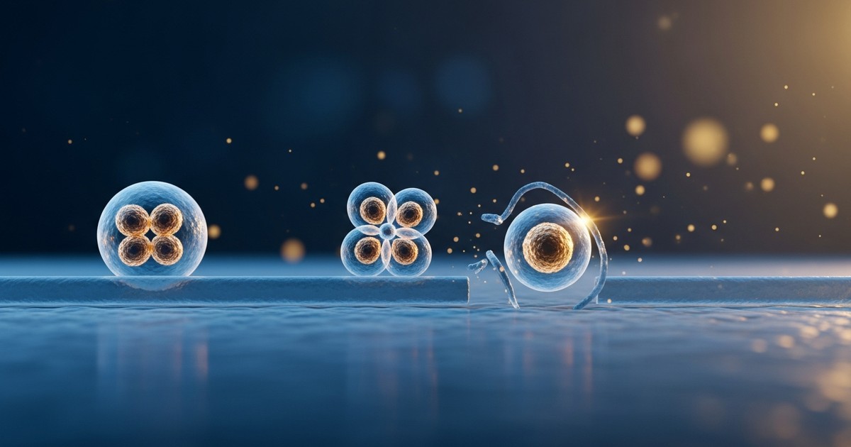
Act Two: Growing in Place — the “Waiting Room” Stage
A cell that has forgotten the rules divides again and again, raising a crowd of copies. The full sequence of preparing and carrying out one division—turning one cell into two—is called the cell cycle. Roughly, the cell cycle is like a wheel turning through four scenes: a phase of preparing to divide, a phase of copying the entire DNA, a phase of checking that copy, and the phase of actually splitting in two. In a healthy cell, the gates we just met halt the wheel here and there, turning it through one careful revolution only when needed. In cancer cells, those gates are broken, so the wheel is hard to stop; it keeps turning. One becomes two, two become four—doubling builds startling momentum in sheer numbers. A test value called Ki-67, which we’ll meet in Vol.5, is essentially a gauge of “how many cells are currently turning that wheel,” and it offers a clue to how forceful this momentum is.
At this stage, though, the cancer is still growing only inside the place where it was born: inside the duct if it began in a duct, inside the lobule if it began there. This state of staying in place is called carcinoma in situ. When it stays within the duct, it is called ductal carcinoma in situ, often shortened to DCIS.
Why does it “stay”? Just outside the duct’s lining cells sits a thin but tough partition called the basement membrane, backed on the inside by sentinel cells called myoepithelial cells. This double wall pens the cancer cells inside the channel. The cancer may be multiplying, but it has not yet left the “waiting room.” This is why carcinoma in situ is also called “stage 0,” and why, handled properly, its outlook is excellent: the wall has not yet been broken. This is the second turning point—inside the wall, or outside it.
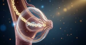
Act Three: Breaking the Wall — the Decisive Line of Invasion
The story turns hard when cancer cells break through that basement membrane and seep outside. This is called invasion. Crossing from in situ (staying in place) to invasive (spreading beyond the wall) is the single most decisive shift in how breast cancer grows.
How is the wall broken? Research shows it is not brute force. The sentinel myoepithelial cells lose their character and can no longer hold the wall up; the basement membrane frays and fragments here and there. At the same time, cancer cells release enzymes that dissolve the surrounding tissue, oozing out through the gaps. Cancer finally sets foot into the stroma—the supporting soil around the ducts and lobules that we met last time.
What happens here is not only that the wall breaks. The cancer cells change character, too. Cells that had been holding hands tightly with their neighbors, lined up neatly in a row, let go of those hands, change shape, and begin to behave like loners able to move one by one. Researchers describe this transformation as epithelial cells changing wardrobe into a more mobile, migratory state; in plain terms, it is a shift from “cells fixed in place” to “cells that can move,” advancing hand in hand with the breaching of the wall. Only cells that have gained the power to move can ooze out through the gaps. The wall weakening and the cells becoming mobile—only when these two come together is the line of invasion crossed.
Whether this “invasion” has happened is judged by a pathologist examining the tissue under a microscope, looking at whether cancer cells lie outside the basement membrane. The distinction between DCIS staying in situ and invasive cancer that has crossed the wall is one of the most important dividing lines in diagnosis and treatment. We’ll examine this line at an expert level later, in Vol.12, “What Pathology Sees.” For now, as a bridge, simply hold onto the biological skeleton: whether or not the wall is crossed divides the outcome.
Act Four: Laying Its Own Plumbing — the Lifeline That Fuels Growth
Even cancer that has invaded the stroma cannot, left alone, grow very large. There is a surprising ceiling here.
Cancer cells, too, need oxygen and nutrients to live, delivered as they seep out of blood vessels into the surrounding cells. But oxygen can only diffuse out from a vessel about 0.1 to 0.2 millimeters. Cells farther than that from the nearest vessel receive no oxygen. Because of this physical limit, a tumor with no blood supply of its own tops out at roughly 1 to 2 millimeters. Its core cells fall into oxygen starvation (hypoxia) and can grow no further. One to two millimeters is so small it cannot be felt by hand and would barely register on imaging. Many cancers may linger, unseen at this tiny size, quietly for a long time.
Here cancer plays a clever hand. The oxygen-starved cells, through sensors that detect low oxygen (chief among them a protein called HIF), release a “summoning signal” called VEGF (vascular endothelial growth factor), calling out to nearby vessels: “send a branch this way.” New vessels then grow toward the tumor mass. This driving of new blood-vessel formation is called angiogenesis. By laying its own supply line, the cancer breaks through the 1–2 millimeter ceiling and grows much larger. This is the third turning point—can it lay the plumbing or not. Only tumors that flip this “angiogenic switch” advance into full-scale growth.
Put the other way around, unless this switch flips, a cancer mass can stay small, marking time. Even among cancers that have crossed the wall, only those able to lay a supply line move ahead—and this is one reason the “speed of growth” varies so widely from one cancer to the next. What’s more, blood vessels built in a hurry by a tumor lack the orderly structure of normal vessels: their walls are full of gaps, they are twisted, and they leak—a misshapen plumbing. This “shoddy construction” later casts a shadow over treatment, making drugs harder to deliver and giving cancer cells openings to slip into the vessels. Why does one cancer summon vessels while another does not? Telling those triggers apart precisely remains, even now, a question at the frontier of research.
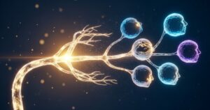
Finale: The Doorway of Metastasis — and Why “Early” Is Decisive
Angiogenesis carries a second, graver meaning. The vessels that grow up to the cancer are supply lines for nutrients—but they are also the boarding platform from which cancer cells set off across the whole body. And, as we just saw, these new vessels have flimsy, gap-riddled walls. For a cancer cell, there is no more convenient “boarding platform” to slip into.
Among invaded cancer cells, some take the “power to move” gained in Act Three a step further, slip through the wall of these new vessels, and board the bloodstream or lymph flow. This step of entering a vessel is called intravasation, and it is precisely the doorway of metastasis. When a cancer cell riding the flow reaches a distant organ—lung, liver, bone, brain—and starts to grow there anew, that is “metastasis.” This is the last and heaviest turning point, the fourth—stay, or set off.
Yet not every cell that sets off builds a new cancer right away. Many cancer cells riding the flow give out along the way, and even those that arrive may not grow at once—they can stay dormant, asleep, for a long time. This capacity to “sleep” is the true nature of one of breast cancer’s hardest features: recurrence years, even decades, later. When and why these dormant cells wake is a major frontier theme we’ll take up in depth in the series’ final volume, Vol.18.
Looking back over the whole story, the reason early detection is so decisive comes clearly into view in the language of biology. “Early” means the story is still in its first half. Found while still in situ before the wall is crossed, or while invasive but still small and before cells have boarded a vessel, the cancer is still in “where it was born and just around it.” It can often be removed completely by surgery, and the heavy treatments that assume a body-wide journey can frequently be avoided. Conversely, once the story has run into its late acts, after cells have already set off, treatment grows far harder. “Early” is not a magic word. It is the biological reality that action is still possible before the turning points are crossed that gives the word its weight.
In Vol.5, we step into the true identity of the protagonist we’ve been lumping together as “the cancer cell.” Though they may look like they grow the same way, breast cancers wear “five faces” with utterly different personalities, and it is that face that decides which drugs work and what the outlook is. Ki-67, which appeared here only as a name, begins to carry meaning there too. The “subtypes” we promised back in Vol.1—at last, we’ll meet them head-on.
My Thought
Tracing how cancer grows as a story brings one quietly reassuring discovery: cancer is not something that “appears fully formed one day,” but the result of crossing several gates, one at a time. That there are gates means there are also chances to stop it midway, and chances to find it. Screening and “awareness” work precisely because this story has several stations along the line.
Stepping in as a writer, the four turning points sketched here—disabling the checkpoint and the self-destruct program, breaching the wall, angiogenesis, and entry into a vessel—line up almost exactly with the targets of modern breast cancer treatment: drugs that re-impose a halt on the dividing wheel, drugs that block angiogenesis, research that cuts off the footholds of metastasis. To understand how cancer grows is to hold a map of why those drugs work.
Yet the story still has a large blank. Why does one carcinoma in situ stay quiet for a lifetime while another breaks the wall and runs wild? We still cannot tell them apart with full confidence. Which gate will be crossed, by whom, and when—predicting that precisely is the hard problem the frontier turns to next. The pathology, diagnosis, and precision medicine this series takes up later are, in essence, an attempt to fill exactly this blank.
Continue the Series
- Vol.1: Breast Cancer Is Not One Disease (the series’ entry point)
- Previous: Vol.3 | Why a Branching “Tree” Turns Cancerous
- Next: Vol.5 | The Three Switches Behind the “Five Faces”
Related Reading
If you leave a comment telling us which cancers or conditions you’d like us to cover next, we’ll prioritize them in future features.

Comments