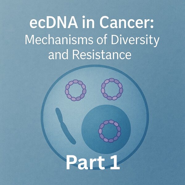
Extrachromosomal DNA (ecDNA) reshapes oncogene dosage, transcription, phenotypes, and spatial organization, thereby accelerating intratumor heterogeneity and therapy resistance. In Part 1, we briefly introduce non-GBM evidence up front and organize the logic of how ecDNA amplifies diversity and adaptation. Part 2 will deep-dive into glioblastoma (GBM).
Key Takeaways (3–5 lines)
- ecDNA lacks centromeres and undergoes unequal segregation, creating cell-to-cell copy-number (dosage) heterogeneity.
- Nuclear ecDNA hubs concentrate transcriptional machinery, enabling rapid, reversible stratification of expression and phenotypes.
- Cross-cancer example: in MYCN ecDNA tumors, high-copy cells → apoptosis-prone vs low-copy cells → senescence-selected, supporting a rational “one-two punch” design.
- The triad of dosage × segregation × hub elevates the speed of tumor adaptation, recurrence, and regional response heterogeneity.
1. What is ecDNA? Core Properties
ecDNA are circular DNA fragments excised from chromosomes. Without centromeres, they fail to segregate evenly during mitosis, yielding copy-number variability across cells. Multiple ecDNAs can cluster into hubs, rewiring enhancer–promoter contacts and concentrating transcriptional apparatus—thus boosting short-term plasticity and long-term evolutionary pace.
2. Set the Stage: Evidence Beyond GBM (brief)
While Part 2 focuses on GBM, ecDNA shapes treatment responses across tumor types. In MYCN-amplified cancers (e.g., neuroblastoma), high-copy ecDNA cells bear replication stress/DNA damage and are apoptosis-prone, whereas low-copy cells enter senescence and are positively selected under therapy—fueling relapse.
The resulting strategy is a rational “one-two punch”: first, doxorubicin eliminates high-copy cells; then, senolytics (e.g., navitoclax) clear residual low-copy senescent cells. Preclinical models, including PDX, indicate reduced regrowth.
3. Why Diversity Accelerates: Dosage × Segregation × Hubs
3.1 Dosage Heterogeneity
ecDNA copy numbers move rapidly, stratifying expression programs and proteomes. Tumors co-host “high-activity, fast-growing, damage-prone” and “low-activity, slow, senescent” subpopulations—priming swift selection games under drug pressure.
3.2 Unequal Segregation & Spatial Asymmetry
Copy-number spikes can arise locally due to biased segregation, seeding spatial heterogeneity that underlies regional response gaps and recurrence-prone niches.
3.3 ecDNA Hubs as “Concentration Fields”
Clustering ecDNA creates high-transcription zones, introducing expression-level differences even at similar copy numbers—broadening phenotypic variance further.
4. Clinical & Therapeutic Implications (Part 1 Overview)
- Dosage-dependent vulnerabilities: high-copy cells are sensitive to DNA-damaging agents; low-copy senescent cells can be targeted by senolytics (e.g., BCL-xL dependence).
- Two-step regimens (“one-two punch”): sequentially combine cytotoxics and senolytics to reduce relapse-capable reservoirs.
- Targeting ecDNA dynamics: conceptual avenues include mitigating unequal segregation or hub formation (early-stage hypotheses).
My Commentary & Outlook (Part 1)
ecDNA are circular DNA elements excised from chromosomes. Because they lack centromeres, they segregate unevenly during mitosis, generating rapid cell-to-cell variability in copy number. That dosage variability cascades into differences in expression, phenotypes, and spatial organization, helping explain core–margin response gaps and the brisk rebound many tumors show after therapy.
Clinically, think in terms of dosage × time. Build an early dosage map via multi-region sampling and imaging fusion; plan a first strike that exploits high-copy vulnerabilities; then perform a senolytic bottom-sweep to clear low-copy senescent/dormant pockets. Stitch these steps together with sequence optimization. In parallel, integrate ecDNA-aware cfDNA metrics and spatial transcriptomics to set explicit thresholds for rebound and pre-define triggers for follow-on therapy. This converts “watchful waiting” into protocolized action tied to quantitative signals.
For readers and teams, keep the mental model compact: a single graphic showing dosage bifurcation → two-step punch, and three closing bullets (unequal segregation, hub-driven transcription, time-sequenced combinations). Don’t treat resistance as a binary state; treat it as a controllable trajectory shaped by dosage dynamics and timing.
Next (Part 2)
We will examine spatial heterogeneity and EGFRvIII emergence in GBM (including the SPECIES model), and translate them into region-aware therapeutic designs.
References
- Extrachromosomal DNA–Driven Oncogene Spatial Heterogeneity and Evolution in Glioblastoma. (PDF)
- Extrachromosomal DNA–Driven Oncogene Dosage Heterogeneity Promotes Rapid Adaptation to Therapy in MYCN-Amplified Cancers. (PDF)
- Extrachromosomal DNA: A Trusted Path to Tumor Heterogeneity. (PDF, Review)
Credits
This article was edited by the Morningglorysciences Team.


Comments