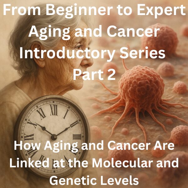
Introduction: Zooming in on the “Inner Structure” of Aging
Aging and cancer share the same cellular clock—and the same failure modes. Mapping the molecular bridge between senescence and tumor biology reveals the design blueprint for a next generation of therapies.
In Part 1, we asked why cancer is often called a “disease of aging” and reviewed how the hallmarks of aging and the hallmarks of cancer overlap. We moved beyond the simple idea that “time passes and mutations accumulate” and instead framed aging as a process that reshapes the cellular and tissue context in which cancer arises.
What you will learn
- Introduction: Zooming in on the “Inner Structure” of Aging
- DNA Damage and Genomic Instability: A Shared Foundation for Aging and Cancer
- Telomeres and Limits to Cell Division: Safety Mechanism and Escape Routes
- Epigenetic Changes and the 3D Genome: Changing How the Switches Work
- Mitochondria, Metabolism, and Protein Quality: Energy and Maintenance
In Part 2, we will look more closely at what that “context” actually consists of at the molecular and genetic levels. Specifically, we will discuss:
- DNA damage and genomic instability
- Telomeres and limits to cell division
- Epigenetic changes and 3D genome architecture
- Mitochondria, metabolism, and protein quality control
- Cellular senescence and the SASP
The scientific literature on each of these topics is vast. Our goal here is not to cover every detail, but to provide a clear conceptual map: what happens in aging cells, how those changes influence cancer risk and behavior, and how to think about these mechanisms without getting lost in jargon and equations.
For specialists, this article is an opportunity to reframe familiar concepts in a broader context. For non-specialists, it is intended as a background that makes it easier to interpret research news and scientific papers on aging and cancer in a structured way.
DNA Damage and Genomic Instability: A Shared Foundation for Aging and Cancer
One of the most obvious common denominators between aging and cancer is DNA damage and the resulting genomic instability. In this section, we will look at how much damage DNA incurs in everyday life, why aging makes that damage more consequential, and how cancer cells exploit this instability.
How Often Is DNA Damaged?
Our DNA is constantly exposed to insults. Ultraviolet light, tobacco smoke and air pollutants, reactive oxygen species (ROS) produced as metabolic byproducts, and background radiation all contribute to a steady stream of DNA lesions. A commonly cited rough estimate is that each cell experiences tens of thousands of DNA damage events per day.
Most of these lesions are quickly and accurately repaired by cellular DNA repair systems. However, repair is not perfect. Some lesions are misrepaired or escape repair altogether, becoming fixed as mutations. In young cells, the balance between damage and repair is biased toward fidelity: repair capacity is relatively high, and the probability that a given lesion becomes a permanent, deleterious mutation is relatively low. As organisms age, this balance gradually shifts.
With aging, multiple DNA repair pathways—base excision repair, mismatch repair, homologous recombination, non-homologous end joining, and others—may decline in efficiency. Repair enzymes may be expressed at lower levels, mitochondrial dysfunction can compromise energy supply for ATP-intensive repair processes, and epigenetic changes may alter how repair factors access damaged sites. The net effect is that DNA repair becomes slower and less accurate, resulting in cumulative genomic instability.
What Changes in Aging: Quantity and Quality of Repair
Aging can be viewed as a slow erosion of DNA repair capacity. It is not simply that fewer lesions are repaired; the fidelity of repair itself may deteriorate. Minor errors that would have been efficiently corrected in youth are more likely to slip through and become fixed as small mutations or structural variants. Over time, this produces a mosaic of genetic changes within tissues.
Some of these changes are functionally neutral. Others alter gene function, signaling pathways, or the integrity of chromosomal regions. In combination with other aging-related changes, this accumulation of genomic “noise” contributes both to tissue dysfunction and to the pool of potentially oncogenic alterations.
Cancer Cells Turn Genomic Instability into a “Weapon”
From the cancer cell’s perspective, genomic instability is a double-edged sword. If genomic chaos is too extreme, the cell cannot survive. But a certain level of instability can be highly advantageous. It provides a steady supply of genetic diversity that allows tumor cell populations to adapt under selective pressures.
When a patient receives chemotherapy or immunotherapy, for example, tumors with high genomic instability may already contain rare subclones that happen to be more drug-resistant or better at evading the immune system. Treatment then selects for these subclones, which expand and drive relapse. In this sense, aging-related deficits in DNA repair and resulting genomic instability not only contribute to cancer initiation but also to the emergence of treatment-resistant disease.
Telomeres and Limits to Cell Division: Safety Mechanism and Escape Routes
Another familiar piece of the aging–cancer puzzle is the telomere. Telomeres are repetitive DNA sequences at chromosome ends, often compared to the plastic tips of shoelaces. They shorten with each cell division due to the mechanics of DNA replication.
Why Do Telomeres Shorten?
DNA polymerases cannot fully replicate the very ends of linear chromosomes. As a result, each round of replication leaves telomeres slightly shorter. When telomeres become critically short, cells activate checkpoints that halt further division or induce apoptosis (programmed cell death). This is one mechanism by which tissues prevent uncontrolled proliferation and reduce the risk of malignant transformation.
Telomere shortening is therefore both a marker and a mechanism of aging. It contributes to the reduced regenerative capacity of tissues over time. However, it also functions as a built-in tumor suppressive barrier.
Telomerase and Replicative Immortality
To bypass this barrier, many cancer cells reactivate telomerase, the enzyme complex that can extend telomeric repeats. In most somatic cells, telomerase activity is low or undetectable. In many tumors, telomerase is upregulated, effectively resetting or lengthening telomeres and allowing repeated divisions without triggering telomere-based checkpoints.
This reactivation of telomerase (or use of alternative lengthening of telomeres, ALT) is a key component of the hallmark of cancer known as replicative immortality. It turns what was intended as a protective limit into a hurdle that only the most “adapted” clones are able to clear.
Integrating Aging, Cancer, and Telomeres
Telomere dynamics sit at the intersection of aging and cancer. Progressive telomere shortening contributes to age-related decline in tissue repair and function and can trigger senescence or cell death in damaged cells. At the same time, if cancer cells manage to reactivate telomerase or ALT, they become capable of near-limitless replication.
In this sense, telomere shortening is both a marker of aging and a tumor suppressive mechanism. When malignant clones learn to circumvent this mechanism, the prior selection pressure effectively “filters out” less fit clones and favors those with robust survival and proliferative mechanisms. Aging and cancer thus interact at the level of telomere biology in a nuanced, context-dependent way.
Epigenetic Changes and the 3D Genome: Changing How the Switches Work
Even when the DNA sequence remains unchanged, the way genes are switched on and off can change dramatically with age. Epigenetics and 3D genome architecture are central to this process.
What Is the Epigenome? “Performance Directions” on the Score
The epigenome refers to regulatory information layered on top of the DNA sequence: DNA methylation, histone modifications, chromatin compaction and relaxation, and more. A common analogy is:
- DNA sequence = the notes in a musical score
- Epigenome = performance directions (tempo, dynamics, instrumentation)
The same score can produce very different music depending on how it is performed. Similarly, cells with identical DNA sequences can adopt very different identities and behaviors depending on their epigenetic state.
Age-Related “Drift” in the Epigenome
With age, epigenetic patterns drift. Typical changes include:
- Loss of methylation in regions that are normally silenced
- Gain of aberrant methylation in promoters of genes that should remain active
- Changes in histone modifications and chromatin compaction that alter which genomic regions are accessible
These changes can lead to inappropriate activation of transposable elements, overexpression of inflammatory genes, and reduced expression of genes involved in stress responses, DNA repair, and antioxidant defense.
The 3D Genome and Cancer: When Spatial Organization Breaks Down
Chromosomes are not randomly packed into the nucleus. They are organized into distinct territories and compartments:
- “A compartments” enriched in active chromatin
- “B compartments” linked to repressed, heterochromatic regions
- Topologically associating domains (TADs) that constrain interactions within regions
This 3D organization helps bring enhancers into contact with their target promoters and keep inappropriate interactions at bay. In cancer, this architecture can be disrupted. Examples include:
- Enhancers that normally regulate one gene become juxtaposed to an oncogene, driving its overexpression
- Tumor suppressor genes are relocated into repressive compartments and become epigenetically silenced
Aging-associated changes in 3D genome organization may make cells more susceptible to this kind of pathological rewiring. Over time, small shifts in nuclear architecture can accumulate, increasing the likelihood that the 3D genome “miswiring” characteristic of cancer will arise.
Mitochondria, Metabolism, and Protein Quality: Energy and Maintenance
Aging and cancer are also tightly linked through energy metabolism and protein quality control—issues of “power supply” and “maintenance” for the cell.
Mitochondrial Dysfunction and Reactive Oxygen Species
Mitochondria generate ATP but also produce reactive oxygen species as byproducts of oxidative phosphorylation. With age, mitochondrial DNA damage accumulates, and mechanisms that control mitochondrial biogenesis, fission/fusion balance, and removal of damaged mitochondria (mitophagy) can deteriorate. The result is a population of mitochondria that produce less ATP and more ROS.
Excess ROS damages DNA, proteins, and lipids, contributing to both aging phenotypes and cancer-promoting changes. At the same time, some tumors rewire their metabolism to rely less on mitochondria and more on glycolysis (the Warburg effect), reflecting a complex interplay between mitochondrial function, proliferation, and survival under stress.
Metabolic Shifts and Cancer: Beyond the Warburg Effect
The Warburg effect—enhanced glycolysis even in the presence of oxygen—has become a classic hallmark of tumor metabolism. It is not simply about inefficient ATP production; it enables rapid provision of biosynthetic precursors needed for growth. Aging cells also undergo metabolic shifts, influenced by insulin/IGF-1 signaling, mTOR, AMPK, NAD+ metabolism, and other pathways.
These nutrient-sensing pathways sit at the crossroads of aging and cancer. Interventions that modulate these pathways—caloric restriction, exercise, drugs targeting mTOR or AMPK—may affect both aging trajectories and cancer risk or behavior. Understanding how metabolic remodeling in aging tissues shapes the metabolic options available to emerging tumor cells is an important frontier.
Breakdown of Protein Quality Control (Proteostasis)
Cells maintain protein homeostasis through a network that includes molecular chaperones, the ubiquitin–proteasome system, and autophagy. These systems identify misfolded or damaged proteins, refold them, or target them for degradation.
With age, proteostasis systems weaken, leading to accumulation of aberrant proteins and reduced capacity to respond to stress. This is well recognized in neurodegenerative diseases, but it also matters in cancer. Proteasome inhibitors, for example, are standard therapies in multiple myeloma. For tumor cells under high proliferative and proteotoxic stress, proteostasis can be both a vulnerability and a source of selective pressure that favors particularly stress-resistant clones.
Cellular Senescence and the SASP: From Defense Mechanism to “Soil”
As discussed in Part 1, cellular senescence is a central process linking aging and cancer. Here we look more closely at its mechanisms and dual roles.
Senescence as a First-Line Tumor Suppressor
When cells detect severe DNA damage, critically short telomeres, or oncogenic stress, they can activate pathways involving p53, p16, and others that drive a stable exit from the cell cycle: cellular senescence. Senescent cells no longer proliferate, but they remain metabolically active and secrete a variety of factors.
In the short term, senescence is strongly tumor-suppressive. It prevents potentially dangerous cells from continuing to divide and acquiring additional mutations. From a tissue-level perspective, it is akin to sacrificing a few compromised cells to preserve overall integrity.
The SASP and Inflammaging
When senescent cells persist in tissues over long periods, their secretory activity—the SASP (senescence-associated secretory phenotype)—becomes a major factor in the tissue microenvironment. SASP includes inflammatory cytokines, chemokines, growth factors, and matrix-degrading enzymes. These molecules can:
- Recruit immune cells to the site
- Remodel the extracellular matrix
- Influence differentiation and proliferation of neighboring cells
The net effect is often a state of chronic, low-grade inflammation (“inflammaging”). This environment can, in some contexts, facilitate the growth and invasion of pre-malignant cells, promote epithelial–mesenchymal transition (EMT), and contribute to immune evasion. At the same time, SASP-driven immune recruitment can help clear senescent cells. The balance between these opposing effects is delicate and context-dependent.
Senolytics and the Possibility of “Targeting Aging” in Cancer Therapy
Recently, senolytics—drugs that selectively kill senescent cells—have attracted attention. In animal models, clearing senescent cells can improve healthspan and, in some cases, enhance responses to cancer therapy. This raises the intriguing possibility that manipulating senescence and SASP could become part of integrated strategies to treat cancer and manage treatment-related toxicities.
Many questions remain: which senescent cell populations should be targeted, at what time points, and in which tissues? Still, the notion that “targeting aging processes” could complement direct anti-tumor approaches is an emerging theme in geroscience oncology. We will return to this topic in the final parts of the Introductory and Expert Series.
The “Molecular Map of Aging” and Cancer Driver Mutations
We can now think of aging as a multidimensional “molecular map” composed of DNA damage, telomere dynamics, epigenetic drift, mitochondrial dysfunction, metabolic shifts, proteostasis breakdown, and senescence. Cancer driver mutations—those in genes like KRAS, EGFR, PIK3CA, TP53, and many others—are superimposed on this map.
The Same Mutation Behaves Differently in Different “Soils”
Large-scale cancer genomics has catalogued driver mutations in numerous tumor types, but clinical experience tells us that tumors with the same driver mutation can behave very differently from patient to patient. Part of this variation may be explained by differences in the aging background of the tissue in which the mutation arises.
For example, consider a mutation in TP53 that occurs in a liver already marked by chronic inflammation and fibrosis versus the same mutation in a relatively young, healthy liver. The subsequent selective pressures, inflammatory milieu, and regenerative dynamics are likely quite different, and so the path from mutation to clinically overt cancer may also differ.
Overlap Between Aging Signatures and Cancer Signatures
Transcriptomic and epigenomic analyses allow us to define “aging signatures” (sets of genes whose expression changes with age in a given tissue) and “cancer signatures” (sets of genes characteristic of particular tumors). Recent work is beginning to map how these signatures overlap.
- In some tissues, aging appears to amplify gene expression patterns that also characterize cancer
- In others, aging may dampen proliferative programs and constrain tumor growth
These organ-specific effects suggest that to assess the risk posed by a particular mutation, we must consider not just the mutation itself but the aging context—how far along the molecular map of aging that tissue has traveled and in which directions.
Conclusion: Viewing Molecular Aging as a Dynamic Background, Not a Static Decline
In Part 2, we explored the molecular and genetic links between aging and cancer through several lenses:
- DNA damage and genomic instability: Aging reduces the speed and fidelity of repair, increasing the diversity of mutations that can serve as fuel for cancer initiation and therapy resistance.
- Telomeres and division limits: Telomere shortening is a tumor suppressive safety mechanism, but cancer cells that reactivate telomerase convert this barrier into a selective filter that favors particularly fit clones.
- Epigenetics and the 3D genome: Age-related drift in epigenetic marks and nuclear architecture alters which genes are on or off, creating conditions that can facilitate oncogenic misregulation.
- Mitochondria, metabolism, and proteostasis: Changes in energy production, oxidative stress, and protein quality control shape both aging phenotypes and the vulnerabilities and adaptation strategies of tumor cells.
- Cellular senescence and the SASP: Senescence is initially tumor-suppressive but can, via SASP and inflammaging, create a microenvironment that promotes tumor growth and invasion.
- The molecular map of aging and driver mutations: The impact of a given driver mutation depends on the aging context of the tissue—its epigenetic, metabolic, inflammatory, and structural state.
The key message is that molecular aging should not be seen as a static “decline” layered on top of an otherwise unchanged system. It is a dynamic process that continually reshapes the background against which cancer originates and evolves. Aging influences not only whether cancer emerges but which cancers emerge, how fast they grow, and how they respond to therapy.
In Part 3, we will move from molecules to systems, focusing on immune aging and the tumor microenvironment. We will examine how inflammaging, immune senescence, and changes in stromal cells interact with the molecular processes discussed here to drive or restrain cancer. These topics bring us closer to clinical questions about immunotherapy, infection risk, and the design of supportive care strategies for older patients with cancer.
My Thoughts
Looking at aging and cancer through the lens of molecular mechanisms highlights the richness and complexity hidden behind the phrase “aging increases cancer risk.” DNA damage, telomeres, epigenetic drift, mitochondrial function, metabolism, proteostasis, and senescence are not independent variables. They form an interconnected network that ultimately determines when, where, and how tumors arise—and how they behave under therapy.
What strikes me is that many mechanisms we talk about as “aging processes” evolved as protective or adaptive responses. DNA repair, telomere-based division limits, metabolic adjustments, and senescence are all, in essence, attempts by cells and tissues to maintain integrity under stress. Problems arise when these systems are pushed beyond their limits by chronic insults and time. The turning point at which protective responses become maladaptive—supporting tumor development or limiting resilience to treatment—may be one of the most important concepts in the aging–cancer interface.
I also find the idea compelling that the same driver mutation can mean something very different in different aging contexts. As precision oncology advances, it will not be enough to read the tumor genome alone. Integrating information about the biological age and aging profile of the host tissue—epigenetic, metabolic, inflammatory, and functional—could lead to more accurate risk assessments and more nuanced treatment choices.
Through this series, my aim is to knit together these molecular insights with immune aging, lifestyle and environmental factors, sex and reproductive aging, and emerging tools like aging clocks and AI-based imaging. If we can establish a shared conceptual language across basic science, clinical practice, policy, and investment, we will be better positioned to design interventions that address both cancer and aging in an integrated way.
This article has been edited by the Morningglorysciences team.
Related Articles


Comments