Introduction: Why “Young” Models Struggle to Capture “Old” Cancer
Throughout this Expert Series we have discussed:
Throughout this Expert Series we have discussed:
What you will learn
- Introduction: Why “Young” Models Struggle to Capture “Old” Cancer
- Why We Need Aging-Aware Cancer Models
- Cell-Based Models: Young vs Senescent Cells
- Organoids and Organ-on-Chip: Moving Closer to Human Tissues
- Mouse Models: Beyond Young Hosts
- Epigenetic clocks and ImAge as tools to visualize aging
- Tissue- and genotype-specific aging profiles
- How lymphoma and cancer therapies can accelerate systemic aging
- Reproductive aging and its links to ovarian, uterine, and breast cancers
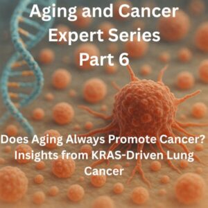
- The KRAS lung cancer model showing that aging can sometimes suppress, rather than promote, tumorigenesis
- Lifestyle, environment, and geography as external drivers of aging × cancer risk
- Geriatric oncology and how to incorporate aging biology into cancer care
A natural question follows: How can we actually model all of this complexity in the lab?
Most preclinical cancer research still relies on:
- Young mice (typically 8–12 weeks old)
- Immortalized cell lines capable of endless division
- Transplanted tumors in immunodeficient hosts
These are, in many ways, models of cancer in a “young and unusually healthy” context. They rarely reflect:
- How tumors behave in aged tissues and microenvironments
- How aged hosts respond to treatments at the systemic level
In this article, using “Challenges and opportunities for modeling aging and cancer” and related work as anchors, we will explore:
- How to model the intersection of aging and cancer across different scales
- The strengths and limitations of cells, organoids, animal models, and human cohorts
- Design principles for building models that are fit for specific scientific and clinical questions
Why We Need Aging-Aware Cancer Models
Most Patients Are Old; Most Models Are Young
The majority of cancer diagnoses occur after age 60. Yet, in preclinical studies:
- Mice are typically the equivalent of human teenagers or young adults
- Cell lines are “ageless,” bypassing replicative limits
This disconnect means that models often miss:
- Age-related changes in tumor microenvironments and host physiology
- Differential toxicity and efficacy of treatments in aged vs young hosts
Aging Is Not Background Noise; It Is Part of Cancer Biology
Aging shapes:
- Tumor-suppressive mechanisms such as DNA repair and apoptosis
- Immune surveillance, inflammation, fibrosis, and angiogenesis
- Metabolic and hormonal networks
It is not a static backdrop but an active participant in tumor initiation, growth, and treatment response. Ignoring aging in models means:
- We are only seeing a subset of the true cancer biology that matters in the clinic.
Cell-Based Models: Young vs Senescent Cells
1) Replicative and Stress-Induced Senescence
At the cellular level, aging can be modeled through:
- Replicative senescence (long-term culture until cells reach their division limit)
- Stress-induced senescence (acute exposure to radiation, DNA-damaging agents, or oxidative stress)
Senescent cells display:
- Stable cell-cycle arrest
- SASP (senescence-associated secretory phenotype)
- Characteristic epigenetic and transcriptomic changes
These features allow us to study how senescent stromal cells influence nearby cancer cells and immune cells.
2) Co-Culture and 3D Systems with Senescent Cells
To better approximate tumor microenvironments, researchers use:
- Co-cultures of senescent fibroblasts with cancer cells
- 3D spheroids or organoids that incorporate senescent components
These systems can reveal how senescent cells alter:
- Cancer cell proliferation, invasion, and drug resistance
- Immune cell function and polarization
3) Limitations: Young Origins and Simplified Architecture
However, many cultured cells come from relatively young donors, and their long-term in vitro history may not fully recapitulate truly aged tissues. Moreover, 2D cultures lack:
- Authentic extracellular matrix, mechanical forces, and nutrient gradients
These limitations constrain how faithfully we can model aged microenvironments at the cell-culture level.
Organoids and Organ-on-Chip: Moving Closer to Human Tissues
1) Patient-Derived Organoids: Bringing Age into the Model
Patient-derived organoids (PDOs) preserve tumor-specific genomes, epigenomes, and drug responses. Crucially, because PDOs are linked to clinical metadata, we can begin to ask:
- How do organoids from older vs younger patients differ in behavior and drug sensitivity?
- How do prior treatments, comorbidities, or reproductive aging status leave molecular “signatures” in organoids?
2) Ovarian and Reproductive Aging in Organoid Models
Studies such as “Reproductive aging leads to many women’s health problems” and “Stress granule clearance mediated by NCOA7 mitigates ovarian aging” highlight molecular mechanisms of ovarian aging, including:
- Stress granule dynamics, autophagy, and mitochondrial function
Future models will likely involve:
- Organoids derived from aged ovaries and reproductive tissues
- Organoids cultured under conditions that mimic aging-related stress
to better understand how reproductive aging intersects with ovarian cancer, infertility, and other conditions.
3) Organ-on-Chip and Multi-Organ Platforms
Microfluidic organ-on-chip systems can recreate aspects of:
- Microcirculation, shear stress, and drug pharmacokinetics
When multiple organs are linked (e.g., liver, adipose tissue, bone marrow), we can study how an aged systemic environment affects:
- Tumor biology
- Treatment toxicity and off-target effects
Such platforms are promising but technically demanding, and integrating aging variables remains an ongoing challenge.
Mouse Models: Beyond Young Hosts
1) Young vs Aged Mice: Same Tumor, Different Behavior
The KRAS-driven lung cancer study “Aging represses oncogenic KRAS-driven lung tumorigenesis…” elegantly demonstrates that:
- The same oncogenic driver (KRAS) yields fewer and smaller tumors in aged mice compared with young mice
- The strength and hierarchy of tumor suppressor pathways change with age
This underscores that:
- The effect of a given driver mutation is strongly modulated by host age and aging state
2) Practical Barriers to Using Aged Mice
Despite their importance, aged mice are underused because they are:
- Costly and time-consuming to maintain
- More prone to spontaneous disease and death, increasing variability
As a result, a pragmatic strategy is to reserve aged mice for:
- Focused questions about treatment toxicity, pharmacokinetics, and immune responses in aged hosts
- Key validation experiments after initial screens in young models
3) Immunodeficient Hosts and PDX: Aged Cancer in Young, Immune-Null Bodies
Patient-derived xenografts (PDX) faithfully mirror many aspects of human tumor biology but are typically grown in:
- Young, immunodeficient mice
This limits their ability to model:
- Aged immune systems
- Aged bone marrow and organ microenvironments
Emerging strategies include:
- Humanized immune-system mice
- PDX in aged or progeroid hosts
but these remain technically challenging and not yet widely adopted.
Human Cohorts and Omics: Real-World Aging × Cancer
1) Longitudinal Cohorts and Three-Dimensional Maps of Aging
Studies like “Physiological aging in three dimensions” and “Tissue-specific impacts of aging and genetics on gene expression patterns in humans” leverage large cohorts and biobanks to map aging across:
- Organ function
- Molecular profiles
- Clinical outcomes
Integrating these data with cancer registries and treatment records enables questions such as:
- Which aging signatures are associated with increased risk of specific cancers?
- Do certain aging patterns predict treatment response or toxicity?
2) ImAge and Single-Cell Spatial Aging Maps
ImAge and related methods infer aging states from single-cell images. Applied to tumor and adjacent tissues, they can reveal:
- How “aged” different cell populations are within the tumor microenvironment
Overlaying this with spatial information about clonal architecture, immune infiltration, fibrosis, and vasculature allows us to ask:
- How do aged microenvironments shape tumor progression and therapy resistance in human tissues?
Design Principles: Fit the Model to the Question
1) Clarify the Question Before “Adding Aging”
Before complicating models with aging variables, it is crucial to ask:
- What exactly do we want to understand or predict?
For example:
- How aged stroma alters drug sensitivity?
- How aged hosts tolerate immunotherapy?
- How reproductive aging influences ovarian cancer initiation?
The answer will dictate which combination of cells, organoids, animal models, and human data is appropriate.
2) Combining “Young vs Old,” “Normal vs Tumor,” and “Single-Organ vs Systemic”
In an ideal world, we would systematically vary:
- Young vs aged hosts and tissues
- Normal vs tumor states
- Single-organ vs multi-organ/systemic models
In reality, resources are limited. A practical compromise is a two-stage strategy:
- Screening stage: use simpler, often young models to identify mechanisms and candidate interventions
- Validation stage: test key findings in aging-aware models (aged mice, aged organoids, human cohorts)
Intervention and Drug Discovery at the Aging × Cancer Interface
1) Geroscience Interventions in Oncology
Geroscience-inspired interventions—mTOR inhibitors, metformin, NAD⁺ boosters, sirtuin modulators, autophagy enhancers—raise questions such as:
- Can targeting aging mechanisms improve cancer prevention or survivorship?
- Can we reduce treatment toxicity or treatment-induced accelerated aging?
Answering these requires models that capture both:
- Aged tumor biology
- Aged host physiology
2) Senolytics and Senomorphics
Senolytics (which eliminate senescent cells) and senomorphics (which modulate SASP) may help:
- Mitigate therapy-induced premature aging
- Reduce recurrence or metastasis driven by senescent microenvironments
However, senescent cells also have beneficial roles in tissue repair and regeneration. We therefore need models that help define:
- Which senescent populations to target
- In which organs and at what time points
Conclusion: No Perfect Model, Only Models that Fit or Misfit the Question
In this ninth article, we have:
- Surveyed cell-based, organoid, animal, and human-cohort approaches to modeling aging and cancer
- Highlighted the limitations of young-only models and the need for aged contexts
- Outlined design principles that align models with specific scientific and clinical questions
- Considered how aging-aware models can support geroscience-inspired interventions in oncology
The key message is that there is no single, perfect model of “real-world aging cancer”. Instead, we must:
- Consciously decide which aspects of aging to include or omit
- Use multiple, complementary models to triangulate on the truth
In the next and final part of this series, we will step back and ask:
- Where might the field of aging and cancer be heading over the next 10–20 years?
- What breakthroughs could emerge in clinical care, public health, and drug development?
My Thoughts
At the intersection of aging and cancer, it is tempting to dream of a universal model that captures everything: young and old, normal and malignant, immune and metabolic, organoids and mice and human data—all in one. In practice, this kind of maximalism often collapses under its own weight. The more we add, the harder it becomes to see which variables are driving which outcomes.
What seems more realistic—and more scientifically honest—is to choose our imperfections deliberately. Use simple young models when they are sufficient, and move to aging-aware systems when the question demands it. Accept that no single experiment will settle a complex issue, and instead design networks of models that cross-check one another from different angles.
For clinicians, the ultimate goal is straightforward: to know which therapies will work, with tolerable risk, for specific patients on specific aging trajectories. The route from bench to bedside will likely involve a mosaic of models rather than a monolith. If aging-aware models can make that mosaic sharper and more predictive, they will have earned their place—not as ends in themselves, but as tools that help patients and clinicians navigate difficult choices with a bit more clarity.
This article has been edited by the Morningglorysciences team.
Related Articles


Comments