# EN|”Suspected” Is Not “Confirmed”: The Work-Up Pathway and Four Lenses for the Wait
After a screening or a clinic visit, hearing the words “we’d like to look more closely” or “you’ll need further tests” can make the mind go blank. In the previous Vol.7, we walked through what happens when you first notice a lump—which clinic to visit, and what is done first. This time we take the next step: the path from being told breast cancer is “suspected” to the moment a diagnosis is confirmed (or ruled out).
Let’s begin with the single most important point. “Suspected” is not “confirmed.” Even when further tests are recommended, most of the time the final answer is “not cancer.” This is not false comfort; as the concrete numbers later will show, it is a fact grounded in probability. This piece aims to convey, accurately, the meaning, the pain, and the time involved in each test—and above all, to support, quietly and with science, how to live through the anxious wait for results. By the time you finish reading, the unknowable weight of the phrase “further testing” should have turned, step by step, into a “procedure” with a clear shape. Vague fear shrinks considerably once it simply has a name.
The Work-Up Moves in Three Stages: Imaging → Needle Biopsy → Pathology
The work-up after breast cancer is suspected stacks, as needed, into three stages. Not everyone goes through all of them; steps are chosen according to the situation. What matters is that this is not “a straight road to surgery,” but a road with many branch points where it can end (“it was benign”). With each step forward, the physician gains more solid information and judges so as not to pile on unnecessary tests.
Stage one: look closely with imaging. The first move is additional imaging. A mammogram may re-image the area of concern with magnification or spot compression, or a breast ultrasound may distinguish whether a lump is a fluid-filled sac (a cyst) or a solid mass. This “cyst versus solid” distinction matters more than one might expect: a fluid-filled sac is benign in the great majority of cases, and on its own is often reassuring. Ultrasound involves no radiation and almost no pain—a gel-coated probe glides over the skin while the examiner observes in real time. You lie on the table for about ten to twenty minutes as the doctor or technologist checks the shape, margins, and blood flow of the finding on the screen. When needed, breast MRI can assess extent and character in still greater detail. MRI is done lying face down, entering a tubular machine, sometimes with a contrast agent, so it takes a little longer (around thirty minutes), but it is not painful. In many cases, this imaging stage concludes that a finding is “a typical benign appearance,” and a person is simply followed over time without ever reaching a biopsy.
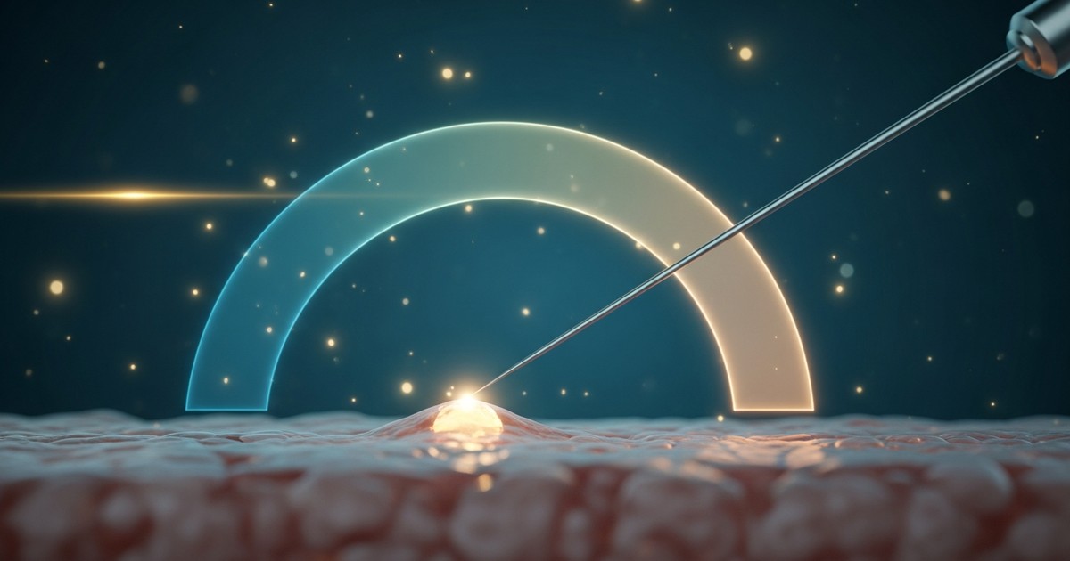
Stage two: take tissue with a needle (needle biopsy). When imaging says “to be safe, let’s confirm the cells or tissue,” a biopsy is done. This is the heart of diagnosis, covered in detail below. It bears emphasizing that proceeding to biopsy does not mean “cancer has been confirmed”; it means “imaging alone cannot settle the question, so we’ll make it certain.”
Stage three: identify the truth with pathology. Examining the sampled tissue under a microscope is pathology. Only here is it confirmed whether cancer cells are present, and if so, what their character is. As a rule, the final diagnosis of breast cancer is made not by the impression on imaging but by this pathology result. The pathologist thinly slices the sampled tissue, stains it, and examines the shape, arrangement, and growth of the cells one section at a time. This is why results take several days to about a week—not because “bad news comes slowest,” but because carefully reading each person’s tissue genuinely takes that time. The deep world of what pathology sees is explored fully in Vol.12, later in this series.
A Needle Biopsy Is Less “Frightening” Than “Quick”—Knowing the Types, Pain, and Time Accurately
“A needle taking tissue” sounds daunting, but the actual burden is often far lighter than imagined. Breast needle biopsies come mainly in these forms. Each balances “how much tissue is obtained” against “burden on the body” differently, and they are chosen according to how the lesion appears.
- Drawing cells with a thin needle (fine-needle aspiration, FNA): a very thin needle on a syringe aspirates cells. It leaves no scar and is quick, but because only “cells” are obtained, the information is limited. It is used, for instance, to drain and check the contents of a cyst.
- Taking tissue with a slightly larger needle (core needle biopsy): the most standard choice when breast cancer is suspected. Under local anesthesia, a needle about the width of a pen refill removes a “core” of tissue. The needle often advances in an instant via a spring mechanism, usually taking three to five cores. Because more tissue is obtained than with FNA, it reveals not only whether cancer is present but also the “character” discussed later.
- Taking ample tissue with suction (vacuum-assisted biopsy, VAB): an evolution of core biopsy that uses suction to collect multiple samples by rotating the needle through a single insertion. It obtains larger and more tissue than core biopsy and suits lesions hard to capture otherwise, such as fine calcifications.
To aim accurately, the needle is guided by a “navigator” suited to how the lesion appears—advancing the needle while watching live ultrasound (ultrasound-guided), or pinpointing the location on mammographic images (stereotactic-guided). For a lesion whose only clue is calcifications, mammography leads the way; for a lesion visible as a mass, ultrasound does—whichever can target it most reliably is chosen.
About pain. All use local anesthesia, so strong pain during the procedure is unusual. There is a stinging, tingling sensation lasting a few seconds when the anesthetic is first injected; once it takes effect, you feel only a sense of “pressure” or “tugging” as tissue is taken, and with core biopsy a spring-loaded “click.” Many people describe it as closer to a “sensation” than pain. About time. The biopsy itself takes from a few minutes up to about thirty, and is usually completed same-day, as an outpatient. Afterward, bruising or mild soreness may appear, but most of it eases with over-the-counter pain relief and settles within a few days. You’ll typically be advised to avoid vigorous activity for a day or two. Understand it as a low-burden test, entirely different from the image of “surgery that cuts open the breast.” Before the procedure, don’t hesitate to confirm any worries—whether blood will be drawn, whether you’ll need someone to accompany you—since that alone can greatly change your peace of mind on the day.
Reading the Probability—Your “True Odds” at the Stage of Suspicion
This is the heart of the piece. When further tests are recommended, the thought that flashes through the mind is “could this already be cancer?” Yet the scenery changes once you correctly read the probability presented at the stage of suspicion.
Breast imaging worldwide uses a shared assessment system called BI-RADS. It is a ruler expressing “how much cancer is suspected” in categories, each tied to an approximate probability of malignancy (cancer). Knowing that a “Category” written on your results sheet is not the physician’s subjective impression but a “language of probability” shared around the world makes the meaning of the numbers far more three-dimensional.
- Categories 1 and 2 (negative / benign): probability of cancer essentially 0%.
- Category 3 (probably benign): probability of cancer 2% or under. Usually this means short-interval follow-up—”let’s look again in six months.” Not biopsying immediately is not an oversight; it is the judgment that “rather than putting a needle into the body for a tiny possibility, it makes more sense to wait a little and confirm there is no change.”
- Category 4 (cannot exclude cancer; biopsy advised): the probability ranges widely, roughly 2–95%, and is further split into 4A, 4B, and 4C. In the most common 4A, the probability is only over 2% to 10%. The next tier, 4B, is over 10% to 50%, and 4C over 50% to under 95%—the probability climbs step by step. In other words, in 4A, the typical case where a biopsy is advised, it is not unusual for around nine out of ten to be benign.
- Category 5 (highly suggestive of cancer): probability of cancer 95% or more.
The key point here is that even within the same “Category 4,” the odds in 4A and 4C are utterly different. That is exactly why, when you hear your result, it is worth going one step further than “it was Category 4” and asking, “within 4, is it A or C?” Even the same “biopsy advised” calls for a different inner stance.
What these numbers tell us is that “a biopsy was advised” does not mean “almost certainly cancer.” Medicine’s caution—”let’s confirm, to be safe”—is what generates many “suspicions,” and most of them land on benign. Being suspected is not a diagnosis. Receive the test not as something meant to frighten you, but as something done to settle the question clearly and free you from needless worry.
Supporting the Wait—Four Concrete Lenses
Even so, the days to a week from test to result can feel like one of the longest stretches of a lifetime. How to spend this “wait”? Here are four lenses—not platitudes, but genuinely useful.
Lens 1: Know that anxiety is a “natural response,” not an “abnormality.” While awaiting results, sleeplessness, an inability to concentrate, runaway worst-case imagining—these are not weakness but the normal workings of a brain facing a threat. Faced with an uncertain threat, the human brain tries to “prepare” by repeatedly rehearsing the worst-case scenario. That itself is a survival function, not a malfunction. Don’t suffer twice by telling yourself “something is wrong with me for being this shaken.” Not making anxiety itself the enemy is the first support.
Lens 2: Translate the probability into “your own words.” Recall the BI-RADS numbers above. If your doctor said “Category 4A, cancer probability around ten percent,” that means “most likely benign—about nine in ten—but we’ll confirm to be safe.” Simply replacing a vague “it might be cancer” with the language of concrete probability shrinks the outline of fear remarkably. If ten people stood in the same situation, about nine of them would hear “it was benign” and go home—picturing it that concretely lets you see where you actually stand. If unsure, you may ask your physician plainly, “What kind of probability are we talking about here?” Asking about the odds is neither selfish nor neurotic; it is simply your right.
Lens 3: Separate out “what you don’t have to decide yet.” Beginning, before results, to weigh treatments, surgery, work, and family makes anxiety swell without limit. Shelve, for now, “the things to think about once results are in.” Resolving that all you can do today is wait is not avoidance but a wise use of time. For the worries circling in your head, it can help to write them on paper and sort them into two piles—”this is for after the results” and “this I can do today.” Seeing your thoughts physically organized quiets the nighttime rumination a little.
Lens 4: Deliberately protect your body and daily rhythm. A light walk, eating and sleeping as usual, saying a word to someone you trust. Obvious as it sounds, keeping the foundation of daily life physically calms thinking that tends to spiral. The person you talk to need not be many. Having even one person to whom you can say “I’m waiting on a test result and I’m a little anxious” greatly lightens the burden of carrying it alone. Conversely, repeating late-night searches and being swallowed by fragmentary information only amplifies fear. Putting distance between yourself and information at night is, in itself, a fine form of self-protection.
How to Tell “Trustworthy Information” Apart
In the waiting, we seek information—and here lies a trap. What surfaces first in a search is not necessarily correct. A few clues help identify trustworthy sources.
- Is the source clear? Public cancer institutions, professional societies, university hospitals, national health-information sites—places where responsibility is plainly located. National cancer societies and major academic centers are good benchmarks.
- Does it avoid absolutes like “guaranteed cure” or “this alone prevents it”? There is no 100% in medicine. Extreme certainty, or fear-driven steering toward a particular product or paid treatment, is a warning sign. The restless urge to “do something while waiting” is precisely the moment when such information can sweep your feet out from under you.
- Does it avoid confusing one person’s story with the general rule? Patient blogs hold great value, but one person’s course is not your own odds. Read the language of averages and probabilities and the language of individual stories from separate drawers. Keep in mind, too, the search bias by which the most vivid, heaviest accounts tend to surface most readily.
- The final anchor is the physician who is looking at your results. Online information helps you prepare emotionally, but only the doctor actually seeing your images and tissue can speak about you. Note your questions and be sure to ask at your next visit. Jotting down what you want to ask beforehand helps you avoid missing anything in the tense setting of a consultation.
Note that this series, including this piece, is a map for understanding the disease correctly—not a source of individual diagnosis or treatment decisions. Final judgments should always be made in dialogue with your treating physician.
In the next Vol.9, we walk further—if you are told “it is breast cancer,” how the heart moves from that day, and how to take the first step. It is a map for reading stage and survival figures through the lens that “an average is not an individual’s fate,” and for deciding calmly. Should you cross beyond suspicion and face the reality of a diagnosis, let’s trace together the path that keeps you from carrying it alone.
My Thought
Speaking with people who have been advised to undergo further testing, I sense that much of their anxiety is really about not-knowing. What will be done, how much it will hurt, what the odds are—because these are unknown, the imagination races toward the worst. That is precisely why carrying the work-up pathway, and a probability ruler like BI-RADS, as “your own words” has practical value.
Going a step deeper, a dilemma of modern medicine lies here. The harder we try to catch breast cancer “early and without misses,” the wider the net of “suspicion” spreads as the price of caution—imposing tests and anxiety even on those who turn out benign. That most BI-RADS 4A findings land on benign is the flip side of how deliberately screening and work-up are designed to “err on the safe side.” The ideal is to refine this net so it lifts up only those who truly need it. The sharpening of diagnostics and AI-assisted image reading are moving in exactly this direction—shrinking the “uncertainty of suspicion”—and that frontier is taken up in Vol.13. While trusting a future in which technology one day makes the anxiety of waiting vanishingly small, for now I hope the solid fact that “suspected is not confirmed” can serve as a quiet talisman.
Continue the Series
- Vol.1: Breast Cancer Is Not One Disease (the series’ entry point)
- Previous: Vol.7 | When You Find a Lump
- Next: Vol.9 | From the Day You Were Told
Related Reading
If you leave a comment telling us which cancers or conditions you’d like us to cover next, we’ll prioritize them in future features.


Comments