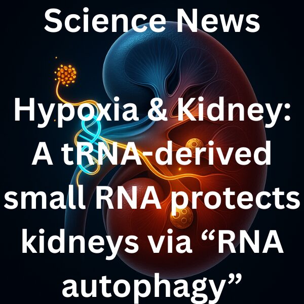
Lead: Kidneys are prone to hypoxia, fueling AKI-to-CKD progression. A 2025 Science study shows that a hypoxia-induced “tRNA-Asp-GTC-3′tDR” safeguards kidney cells by maintaining autophagic flux through RNA autophagy. This adds a new layer to the classic HIF/metabolic view: tDR → PUS7 sequestration → reduced pseudouridylation of histone mRNAs → activation of RNA autophagy.
TOC
Key Takeaways (3 bullets)
- Hypoxia-responsive 3′tDR rises in kidney and preserves autophagy, limiting injury, inflammation, and fibrosis.
- 3′tDR forms G-quadruplexes to sequester PUS7, reducing pseudouridylation of histone mRNAs and triggering RNA autophagy.
- In mouse models, 3′tDR mimics are renoprotective, whereas ASO silencing worsens disease—supporting nucleic-acid therapeutic avenues.
Background: Why kidney hypoxia matters
- Medullary pO2 is physiologically low, while proximal tubular Na reabsorption drives oxygen demand.
- Proteinuria, diabetes (SGLT2 overactivity), anemia, and capillary rarefaction skew supply–demand, leading to chronic hypoxia → inflammation/fibrosis → GFR decline.
- HIF activation is adaptive acutely but can promote fibrosis when chronic.
New Finding (Science 2025): tDR-driven RNA autophagy protects the kidney
1) What was discovered?
- Under hypoxia/renal stress, tRNA-Asp-GTC-3′tDR increases prominently and is basally high in primary kidney cells.
- 3′tDR is necessary and sufficient to sustain autophagic flux; 5′tDR shows limited effect. Silencing 3′tDR reduces autophagy and increases cell death.
2) Mechanism
- 3′tDR assembles G-quadruplexes (G4) via oligo-G motifs and binds PUS7, functionally “parking” the enzyme.
- Reduced PUS7 activity lowers pseudouridylation of histone mRNAs, routing them to the autophagosome–lysosome pathway and activating RNA autophagy, thereby sustaining cellular homeostasis under stress.
3) In vivo & Translatability
- 3′tDR rises early in multiple murine kidney disease models and human tissues.
- LNA-ASO knockdown exacerbates injury, inflammation, and fibrosis.
- Delivery of synthetic 3′tDR mimics via polymer nanoparticles preserves autophagy and lowers injury/fibrosis markers.
Clinical Implications
- Lower demand: SGLT2 inhibitors, RAAS blockade, salt restriction, and BP/Glucose control.
- Raise supply: Treat renal anemia (iron/ESA/HIF-PHI with cautious targets), manage OSA; lifestyle to improve endothelial health.
- New layer: Kidney-targeted tDR therapy could bridge upstream oxygen economy with downstream autophagy homeostasis.
Development Optics (Where to play)
- Modality: 3′tDR mimics retaining PUS7-binding and G4 stability with nuclease resistance.
- Delivery: Kidney-tropic nanoparticles (size/charge/ligands) for proximal tubule/medulla targeting.
- Indications: AKI (peri-operative/contrast, sepsis), diabetic kidney disease, post-transplant IRI.
- Combinations: Complement with SGLT2i/RAAS to optimize dose windows and safety.
- Biomarkers: Urine/serum tDR, autophagy readouts, and functional MRI to stratify responders.
- Safety: Monitor for excessive autophagy and off-target effects on RNA modification networks.
Future Therapeutics: My Take
- Nucleic-acid lead: Engineer 3′tDR mimics preserving the PUS7-binding epitope and G4 motifs; establish in-kidney exposure and autophagic-flux PD anchors.
- Targeted delivery: Use sugar/peptide ligands for receptor-mediated uptake in proximal tubules; scale polymer NP platforms with QbD.
- Time-windowed designs: Prophylaxis in AKI vs. disease-modifying therapy in CKD.
- Combo logic: Align oxygen economy (SGLT2i/RAAS) with autophagy homeostasis (tDR).
- Dx–Rx co-development: Build a companion diagnostic around urinary tDR and histone mRNA modification status.
References
- Li G, Sun L, Xin C, et al. A hypoxia-responsive tRNA-derived small RNA confers renal protection through RNA autophagy. Science. 2025;389(6763).
This article was edited by Morningglorysciences.


Comments